Every Diagnosis Starts With a Perfect Section
Explore the technology inside our tissue processing systems.
The Blade-to-Block Interface Where Diagnosis Is Won or Lost
Every artifact in a pathology report — compression, chatter, thickness variation — originates at the cutting edge. Specimen's rotary microtome chassis is machined from a single billet of 316L stainless steel, eliminating the resonance nodes that cause ribbon irregularities in cast-frame competitors.
Microtome Specifications — SM-7 Series
Cross-Section Diagram — Blade-Block Interface
Hover hotspots to inspect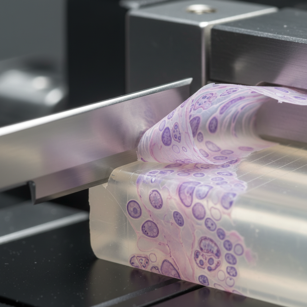
Tungsten-carbide blade edge
3 µm clearance angle
Specimen chuck clamp
±0.1 µm repeatability
Paraffin ribbon output
1–60 µm section range
Coarse advance mechanism
1 µm fine feed resolution
Blade Advance Sequence
Sectioning Sequence — What Happens in 0.3 Seconds
- 01Specimen advance — Motorized coarse feed positions block face 3 µm below blade plane.
- 02Blade engagement — Tungsten-carbide edge contacts paraffin at 3° clearance angle, shearing cleanly without compression.
- 03Ribbon formation — Section exits blade face and adheres to previous section via paraffin surface tension — forming a continuous ribbon.
- 04Anti-chatter dampening — Pneumatic isolation base absorbs resonance; chassis geometry prevents harmonic feedback into the cutting plane.
See the full sectioning quality data
The Sectioning Quality Guide includes 14 pages of ribbon photography, artifact taxonomy, and blade-selection charts for every tissue type.
Reagent Carousel Engineering That Eliminates Carry-Over
Specimen's TP-12 tissue processor runs a 12-station reagent carousel with individual station sensors. Each bath is monitored for temperature (±0.5°C), reagent level, and contamination index — displayed on a 10.1" capacitive touchscreen. When xylene carry-over into paraffin exceeds 0.3%, the system flags the station before the next cassette run.
12
stations
Independent temp control
±0.5°C
accuracy
Per-bath temperature
0.3%
threshold
Xylene carry-over alert
300
cassettes
Per 12-hour overnight run
TP-12 Reagent Station Map — Live Sequence
TP-12
12
STATIONS
Carousel completes one cycle per 30-minute protocol step
Specimen Journey — Grossing to Embedding
Grossing
Fixation
70% EtOH
95% EtOH
100% EtOH
Xylene
Paraffin
Embedding
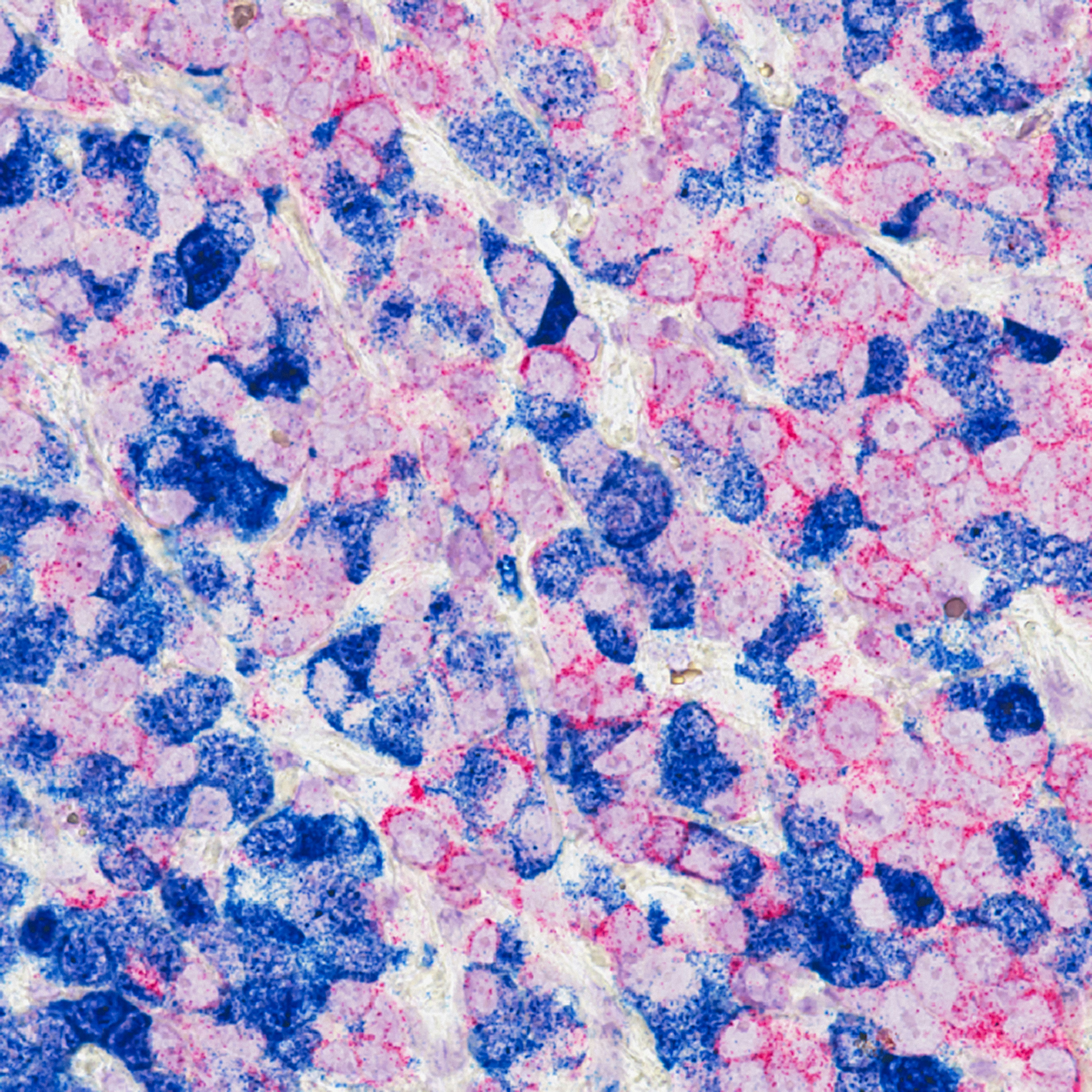
H&E section at 40× — 3 µm thickness, SM-7 microtome
Section Thickness Determines Stain Intensity. We Hold 3 µm.
Stain intensity in H&E and IHC protocols is directly proportional to section thickness. A 5 µm section where a 3 µm was ordered produces over-stained nuclei that obscure Ki-67 scoring. Specimen's SM-7 holds 3 µm ±0.2 µm across a full 60-cassette run — verified by laser profilometry at the factory.
Intraoperative Diagnosis in Under 8 Minutes, Consistently
Frozen section turnaround time is not just a convenience metric — it determines how long a patient remains under general anesthesia. Specimen's CS-3 cryostat reaches −35°C in 12 minutes from ambient, maintains ±0.5°C cabinet uniformity, and produces artifact-free sections from fresh surgical specimens without pre-cooling.
12 min
To operating temp from ambient
±0.5°C
Cabinet temperature uniformity
8 min
Biopsy-to-report turnaround
−35°C
Minimum cabinet temperature
- UV-C decontamination cycle between specimens — 90-second automated protocol
- Anti-ice blade warming strip — eliminates freeze artifact at tissue-blade interface
- Motorized specimen disc with 6-position capacity for concurrent biopsy processing
- Integrated defrost cycle with specimen protection lock-out
Intraoperative Frozen Section — 8-Minute Protocol
- 0:00Specimen received from OR
- 0:30Grossing and orientation
- 1:30OCT embedding, cryo chuck mount
- 2:30First sections cut at 5 µm
- 4:00Rapid H&E (60-second protocol)
- 6:00Slide coverslipped and reviewed
- 7:45Pathologist report to surgeon
Every Instrument Speaks Your LIS Protocol
Specimen instruments ship with HL7 v2.5 and FHIR R4 integration modules pre-installed. The SM-7, TP-12, and CS-3 each expose a RESTful API for programmatic control, allowing your LIS to trigger processing runs, query instrument status, and pull QC logs without manual data entry.
Bidirectional LIS Interface
HL7 + FHIRHL7 v2.5 and FHIR R4 — accession numbers flow from LIS to instrument; run data flows back automatically.
REST API Access
REST APIFull instrument control and status query via documented REST endpoints. Python and R SDK available.
QC Log Automation
ISO 15189Every run generates a tamper-evident QC log in PDF/A format, automatically filed to your document management system.
Multi-Site Dashboard
Multi-siteReference laboratories operating 3+ sites view all instrument status, reagent levels, and throughput from a single web dashboard.
Throughput Comparison — Specimen vs. Industry Average
SM-7
Rotary Microtome
TP-12
Tissue Processor
CS-3
Cryostat
Compatible LIS / LIMS Platforms
"We replaced two aging Leica microtomes with the SM-7 last quarter. Ribbon consistency improved immediately — our histotechs noticed within the first block. The 3 µm hold across a full cassette run is exactly what the spec sheet claims."
Dr. Patricia Holloway
Laboratory Director
Northwell Health Pathology
"The TP-12's xylene carry-over alert has already caught two reagent contamination events we would have missed. That's two patient cases that didn't have to be re-processed. That's the ROI conversation I needed for my department head."
Marcus Okonkwo, HT(ASCP)
Chief Histotechnologist
Cleveland Clinic Main Campus
"Comparing throughput specs across seven vendors took three weeks. Specimen was the only manufacturer that provided laser profilometry certificates for section thickness verification. That level of documentation closes procurement reviews faster."
Sandra Fitzpatrick
Capital Equipment Procurement
Quest Diagnostics — Northeast Region
The Sectioning Quality Guide — 14 Pages of Internal Engineering
What's inside the guide competitors don't publish:
- Ribbon photography at 1, 3, 5, and 10 µm — same tissue block, same staining protocol
- Artifact taxonomy: chatter, compression, venetian blind, score marks — causes and fixes
- Blade selection matrix for 23 tissue types (bone, brain, breast, GI, skin…)
- Section thickness CV comparison: SM-7 vs. four competitor models (laser profilometry data)
- Paraffin formulation guide for optimal ribbon formation at each section thickness
Also available: Blade Sample Kit
Request a physical kit with 5 blade types, 3 paraffin samples, and a 10-slide comparison set — shipped to your lab within 5 business days.
Request a Blade Sample KitDownload Free — No Sales Call
Three fields. Immediate PDF delivery to your work email.
14-page PDF · Immediate delivery · No sales follow-up
4,200+
Instruments in active clinical use
99.2%
Instrument uptime (12-month rolling avg)
3 µm
Guaranteed section thickness accuracy
8 min
Frozen section turnaround, CS-3 cryostat